This is dependent on the stage of disease and needs to be discussed with your doctor.
Your treatment may include the following modalities:
Radical Orchidectomy
A radical orchidectomy should be performed to permit histologic evaluation of the primary tumour and to provide local tumour control.
Chemotherapy
Chemotherapy is used when the tumour has high risk features or when there is evidence of spread.
Radiotherapy
High dose X-ray is used for specific tumour types when there is evidence of spread.
Follow-up after treatment for testicular cancer
After receiving treatment, patients are advised to do regular testicular self-examination and for close follow-up with your treating urologist.
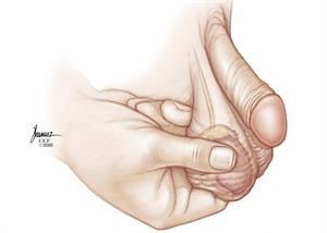
How to do testicular self-examination
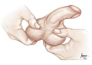
Step 1
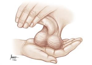
Step 2
Step 3
Response to treatment is assessed after the initial induction cycle by repeat imagining and re-evaluation of tumors markers.